- Have Revised
Bio-signals
All types of signals that can be measured in relation with the human body and its physiology, we will look a bit at the classifications of biosignals
Electrical
Electrical biosignals usually measure biopotentials and impedance, for example:
- Electroenecephalogram (EEG) - measures electrical activity of brain, electrodes placed on scalp and record tiny electrical impulses produced by neural activity
- Electrocardiogram (ECG) - records the electrical signals generated by the heart as it beats, electrodes placed on the skin of chest, arms, legs (usually 12 leads)
- Electromyogram (EMG) - measures electrical activity of muscles during rest and contraction, again electrodes on skin above muscle
- Galvanic skin response (GSR) - measures changes in electrical conductance of the skin, which varies with moisture level, can measure changes in sweat gland activity influenced by the ANS
- Electrical Impedance tomography (EIT) - A medical imaging technique that produces images based on electrical conductivity distribution on body, small electrical currents are applied via electrodes around region of interest and voltage responses are measured
Chemical
This includes things like concentration of things, such as blood glucose
Mechanical
This is mostly measured externally but can also be internal including things such as:
- Pressure (blood for example)
- Temperature (body for example)
- Flow
- Velocity
- Acceleration
- Force
- etc
Imaging
This includes methods such as ultra-sound, X-ray, MRI, etc
Electric signals in living organisms: where do they come from?
More or less all cells have electrical potential -> For some cells this is essential for functionality of those cells.
Structural and functional organisation of the human body
Firstly lets cover the basic fundamental organisation of biological organisms
Cells - fundamental units of a living body, the smallest anatomical and physiological unit of the human body that can live and reproduce on its own
Tissues - a group of cells (and surrounding substances) that function together to perform some function(s). The four primary types of tissue:
- muscle
- nervous
- epithelial (skin, intestines)
- connective (e.g bone, cartilage, ligaments, tendons, etc)
Organs - combination of tissues that perform complex tasks (e.g kidneys, liver, heart, etc)
Systems - there are 11 major organ systems: muscular, skeletal, nervous, respiratory, circulatory, reporductive, digistive, urinary, endocrine, lymphatic, integumentary (skin, hair, nails, etc)
Cells and the cell membrane
The cell membrane is fundamental to the existence of living organisms as we know them. The idea that cells can regulate and compartmentalise themselves and separate themselves from their environment while retaining a structural integrity enables much of the functionality we attribute to living organisms.
The formation of cells and the cell membrane is spontaneous. This is because of phospholipids, which spontaneously form a bilayer when in an aqueous environment.
What are phospholipids?
Phospholipids are a type of lipid molecule that consists of 3 main components:
- Glycerol Backbone: The core of the molecule connecting the different marts
- Phosphate Group: Attached to the glycerol backbone, making one end of the molecule hydrophilic (water-loving)
- Fatty Acid Tails: Two long chains of hydrocarbons that are hydrophobic (water repelling, or not attracting)
Each phospholipid has a polar (hydrophillic) head that includes the phosphate group and a nonpolar (hydrophobic) tail made of fatty acids. This dual nature is curcial for the formation of the cell membrane.
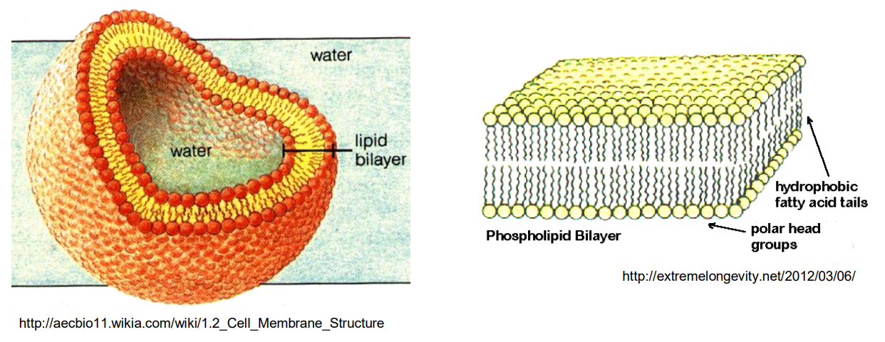
The polar heads and nonpolar tails create a dual nature or amphipathic property, causing the membrane to have very high impedance (MΩ). This provides cells with a selective permeability, only allowing certain molecules to pass through and reject others:
- Oxygen ✅
- Carbon Dioxide ✅
- Ethanol ✅
- water-soluble molecules ❌
- ions ❌
- water itself ❌
✅ = can pass via passive diffusion ❌ = cannot pass via passive diffusion
Therefore specific transport protiens are required in order to allow for useful ions to enter the cell, these take the form of transport channels / enzymes
Potassium and Sodium Ion Channels (Na/K Pump)
Ion channels are proteins embedded in the cell membrane that create pathways for specific ions, such as sodium, potassium, calcium and chlorine to move in and out of cells.
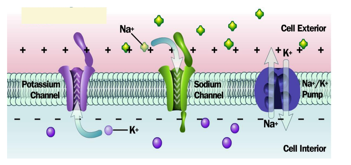
These channels (as seen here) are highly selective, typically only choosing one type of ion to pass through them and can either be gated or leak channels.
So how do ion channels generate currents?
Resting Membrane Potential
Cells (especially neurons and muscle cells) maintain a difference in electric potential between the inside and outside of the cell membrane called the resting membrane potential.
This potential exists because of differences in ion concentrations across the membrane and the selective permeability of the membrane caused by the ion pumps
Ion Movement
Ion channels allow specific ions to diffuse (high to low concentration) toward their opposite charges, e.g sodium ions (+ve charged) will naturally move towards the negative interior of a cell when channels open
When an ion channel opens, ions can move across the cell membrane, this movement of charged particles creates a current
Types of Ion Channels
Voltage-gated channels: These open in response to changes in membrane potential. When a specific voltage threshold is reached, the channels open, allowing ions to flow and create a current.
Ligand-gated channels: These open when a specific molecule (ligand), such as a neurotransmitter, binds to them, allowing ions to flow and generate current.
The opening and closing of these channels are tightly regulated, which in turn controls the timing and amplitude of the current produced.
Transport Processes
What forces things to move?
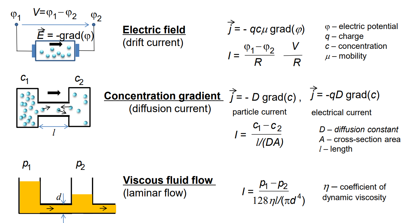
This brings us to the Nernst equation (see here), which relates the concentration gradient of an ion across a membrane to the electric potential at which that ion will be at equilibrium, deriving from:
We get the following equation:
Where:
- is the equilibrium potential for the ion, in volts (V)
- is the universal gas constant, equal to 8.314 J/(mol * K)
- is the temperature, in Kelvin (K)
- is the valence (charge) of the ion
- is the Faraday constant which is approximately 96’485 C/mol
- and represent the concentrations* of the ion inside and outside the cell
Neural Signalling and Communication
Let’s discuss how what we’ve now learnt relates to how the cells in our brain communicate with each other
The Neuron
A diagram of a neuron can be seen here
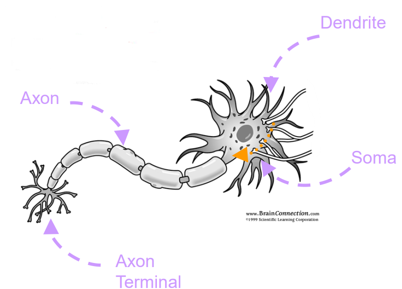
Let’s focus on the most important parts:
Axon - The axon is a long, thin extension of the neuron that carries electrical impulses (action potentials) away from the soma (cell body) toward the axon terminals.
Axon Terminal - The axon terminal, also called the synaptic terminal or terminal bouton, is the endpoint of the axon where the neuron communicates with other cells.
Dendrite - Dendrites are branch-like extensions of the neuron that receive signals (typically in the form of neurotransmitters) from other neurons.
Soma - The soma, or cell body, is the main part of the neuron that contains the nucleus and other organelles.
Transmission
In order to explain how neural signalling works we will look at a very important graph. This describes the action potential of a neuron, which is an electric signal that travels along the axon of a neuron.
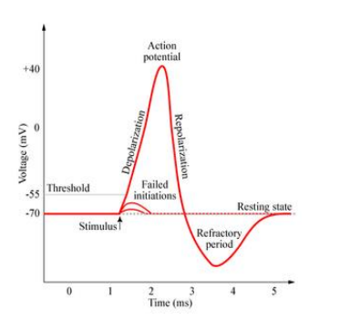
Resting Membrane Potential
- Before an action potential can occur, a neuron maintains a resting membrane potential of approximately -70 mV
- This is maintained due to potassium leak channels, allowing potassium ions to move out of the neuron and sodium-potassium pumps which actively transports 3 sodium ions out of the cell and 2 potassium ions in using ATP
Depolarisation
- The action potential is triggered when the membrane potential of a neuron becomes more positive, reaching a threshold of about -50 mV
- This is usually caused by stimulus from a neighbouring cell
- This causes the voltage-gated sodium channels to open, causing sodium ions to rush into the cell
- This causes the inside of the cell to become much more +ve (as Na+), resulting in depolarisation causing a spike of up to +40 mV
Peak
- At the peak of the action potential the voltage-gated sodium channels start to inactivate, preventing further influx of sodium ions
Repolarisation
- To bring the membrane potential back down, voltage-gated potassium channels open, these open slower than the sodium channels and therefore activate just as the sodium channels inactivate.
- As potassium ions now leave the cell, it causes the membrane potential to become more negative (as K+)
Refractory Period / Hyperpolarisation
- The potassium channels do not close immediately after reaching the resting potential, as a result the membrane potential often becomes more negative than the resting potential
- And after it returns to the resting membrane potential
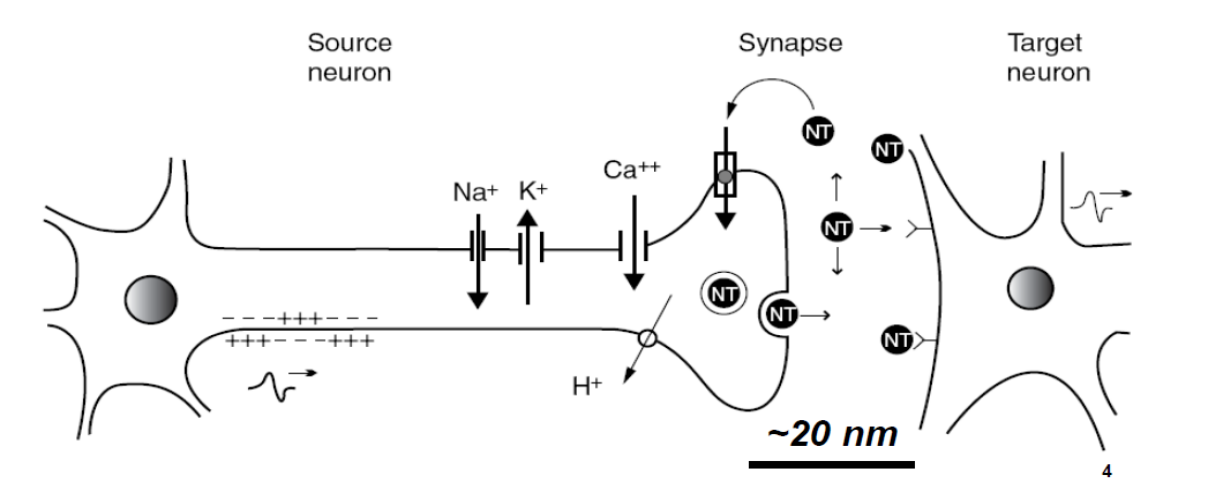
Synapse
- Once the action potential has travelled along the axon and reaches the presynaptic terminal a similar process occurs
- depolarisation causes voltage-gated calcium channels to activate
- This in turn triggers the release of neurotransmitters allowing for the synaptic current to occur (which is a chemical process)
The Nervous System
Zooming out now we can see how this kind of behavior can impact our whole body and system.
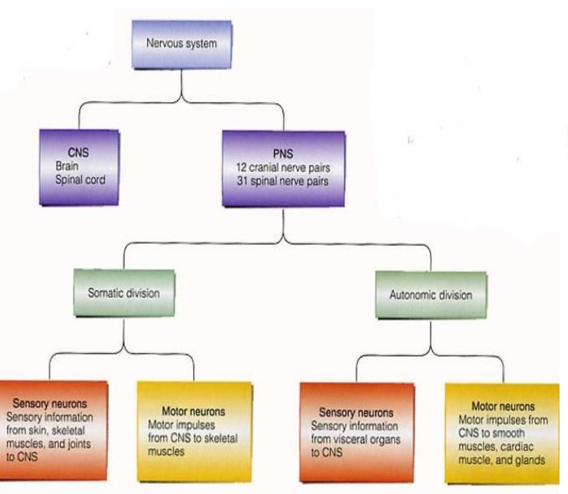
From this image we can see that the Nervous System, dictated by signals and current that we previously discussed, is responsible for A LOT. It is divided between the CNS and PNS and takes up the responsibilities of:
- Sensing the Environment: It detects changes inside and outside the body using sensory receptors (e.g., light, sound, temperature, pressure).
- Processing Information: It interprets and integrates sensory information in the brain to make sense of what’s happening.
- Initiating Responses: It sends signals to muscles and glands to respond to sensory input. This includes voluntary actions, like moving an arm, and involuntary actions, like increasing heart rate.
You can see how important electronics can be therefore with neurological disorders
Typical Biopotentials (EEG, ECG, etc)
EEG measures the brain’s electrical activity from the scalp from the activity of billions of neurons. ECG measures galvanically the electrical activity of the heart.
We can see how these signals can range:
| Signal | Amplitude (mV) | Bandwidth (Hz) |
|---|---|---|
| EEG | 0.001-0.01 | 0.5-40 |
| ECG | 1-5 | 0.05-100 |
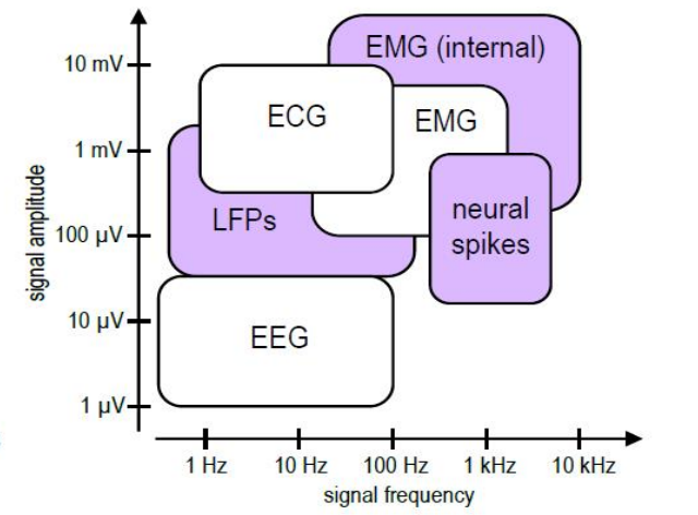
And obviously these can range due to a number of factors
Summary
- The body generates a lot of electrical/physical/chemical signals which give off important information we need to detect to either help monitor, diagnose or treat a medical condition.
- These originate from chemical, bio-physical processes.
- Electronic instrumentation is vital in detecting these signals.
- Proper interfacing with signals of biological origin is still a very challenging task !